The blood-circulating organs are the heart and the blood-vessels. These latter are of three kinds,—the arteries, which carry the blood from the heart to the tissues of the body; the veins, which return the blood from the tissues to the heart; and the capillaries, which are very fine vessels uniting the ends of veins and arteries. They end the arteries, and then gather into larger and larger trunks, thus forming veins. As the veins approach the heart, they form larger trunks, until at last two large veins are formed,—the “descending” and the “ascending” vena cava.
Arteries have firm, elastic walls, and retain their shape, full or empty. They have three layers of connective and muscular tissue. Veins have thinner, less rigid walls. They can hold double the quantity of blood that the arteries do. In the veins are valves, which allow the blood to flow but one way—toward the heart.
The heart is a hollow muscle about the size of one’s fist; conical in shape, placed between the fifth and sixth ribs and the lungs, which nearly cover it. The heart is inclosed in a sac, called the “pericardium.” In this is a fluid secretion, in which the heart moves easily, and by which it is protected from shock. The heart covering is attached to the central tendon of the diaphragm. The weight of the heart is from eight to twelve ounces. A perpendicular muscular partition divides the heart into two halves,—right and left,—and these are again divided into an upper and a lower portion,—four cavities in all. (See diagram.) These cavities are lined with a delicate membrane.
The upper cavities are called the right and left auricles, and the lower cavities are called the right and left ventricles. Each cavity holds about six ounces of blood. The walls of the ventricles are much thicker than those of the auricles; and the wall of the left ventricle is also much thicker than that of the right.
Each ventricle has two openings—one leading to the auricle on the same side, and the other to an artery. The right ventricle connects with the pulmonary artery, which leads to the lungs; and the left ventricle connects with the aorta, the largest artery in the body. The right auricle has two openings in its outer wall, through which the two largest veins in the body (vena cava, ascending and descending) empty the blood into the heart. The left auricle has four openings, which receive the blood from four pulmonary veins, two from each lung.
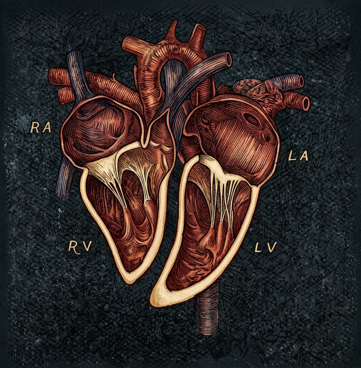
The two openings of the ventricles are guarded by valves, which allow the blood to pass in but one direction. These valves are delicate curtains of membrane. The valve on the right side (between the right auricle and the right ventricle) has three curtains, and is called the “tricuspid valve;” that on the left side has two curtains, and is known as the “mitral valve.” There are also the aortic and pulmonary valves, which are called the “semilunar valves.” These allow blood to flow from the auricles to the ventricles and into the arteries, but not in the opposite direction.
In our next notice we will see how the blood flows through the body, and will study especially a diagram showing the action of the heart in reference to the circulation.
MRS. M. D. MCKEE.